What is the arrow pointing to in image A. Ligamentum Teres B. Ligamentum Venosum C. Main Lobar Fissure D. Falciform Ligament
The Correct Answer and Explanation is:
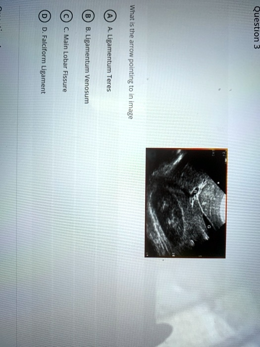
The correct answer is: C. Main Lobar Fissure
Explanation:
In the ultrasound image provided, the arrow is pointing to an echogenic (bright) line that stretches between the gallbladder and the portal vein. This echogenic line is characteristic of the main lobar fissure (MLF).
The main lobar fissure is a key anatomical landmark in liver ultrasound. It is a hyperechoic line (appears bright on ultrasound) that runs between the gallbladder fossa and the right portal vein. It divides the right and left lobes of the liver and provides a reliable guide for locating the gallbladder neck and the porta hepatis.
Radiologists and sonographers often use the main lobar fissure to:
- Identify the gallbladder in challenging cases (especially when it’s contracted or surrounded by bowel gas).
- Distinguish between liver lobes for segmental anatomy.
- Aid in procedures like cholecystostomy or liver biopsy planning.
Let’s look at the other options and why they are incorrect:
- A. Ligamentum Teres: This is a remnant of the fetal umbilical vein and is located within the left lobe of the liver. It runs in the left intersegmental fissure and would not be in line between the gallbladder and the portal vein.
- B. Ligamentum Venosum: This is a remnant of the fetal ductus venosus. It is located more posteriorly and medially within the left lobe and is not in the typical location shown.
- D. Falciform Ligament: This ligament contains the ligamentum teres and connects the anterior abdominal wall to the liver. It does not appear as a bright line running toward the gallbladder on ultrasound.
Therefore, based on the position and appearance of the echogenic line in the image, it is best identified as the Main Lobar Fissure.