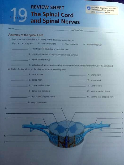
REVIEW SHEET 19 The Spinal Cord EXERCISE and Spinal Nerves Lab Time/Date Name Anatomy of the Spinal Cord 1. Match each anatomical term in the key to the descriptions given below Key: a. cauda equina b. conus medullaris c. filum terminale d. foramen magnum 1. most superior boundary of the spinal cord 2. meningeal extension beyond the spinal cord terminus 3. spinal cord terminus 4. collection of spinal nerves traveling in the vertebral canal below the terminus of the spinal cord 2. Match the key letters on the diagram with the following terms 1. central canal 7. lateral horn 2. dorsal horn 8. spinal nerve 3. dorsal median sulcus 9. ventral horn 4. dorsal root ganglion 10. ventral median fissure 5. dorsal root of spinal nerve 11. ventral root of spinal nerve 6. gray commissure
The Correct Answer and Explanation is:
Of course! Here are the correct answers for the review sheet on the spinal cord and spinal nerves, along with a detailed explanation.
Correct Answers
Part 1: Match each anatomical term in the key to the descriptions given below.
- d. foramen magnum
- c. filum terminale
- b. conus medullaris
- a. cauda equina
Part 2: Match the key letters on the diagram with the following terms.
- central canal: j
- dorsal horn: k
- dorsal median sulcus: l
- dorsal root ganglion: a
- dorsal root of spinal nerve: c
- gray commissure: i
- lateral horn: h
- spinal nerve: b
- ventral horn: e
- ventral median fissure: f
- ventral root of spinal nerve: d
Explanation
This exercise covers the major anatomical features of the spinal cord, from its overall structure within the vertebral column to the details of its cross-section.
Part 1: Gross Anatomy of the Spinal Cord
The answers for the first section relate to the macroscopic structures of the spinal cord. The foramen magnum (d) is the large opening at the base of the skull through which the brainstem connects to the spinal cord, marking its most superior boundary. The spinal cord itself tapers to an end around the first or second lumbar vertebra in a structure called the conus medullaris (b), which is the spinal cord terminus. Extending from the conus medullaris is the filum terminale (c), a fibrous extension of the pia mater that anchors the spinal cord to the coccyx. Because the spinal cord is shorter than the vertebral column, the lumbar and sacral nerve roots must travel downward before exiting. This collection of descending nerve roots is called the cauda equina (a), named for its resemblance to a horse’s tail.
Part 2: Cross-Sectional Anatomy of the Spinal Cord
The second section identifies structures in a cross-section. The diagram’s orientation can be determined by key landmarks. The posterior or dorsal side features the shallow dorsal median sulcus (l), while the anterior or ventral side has the deeper ventral median fissure (f).
The gray matter, shaped like a butterfly, contains neuron cell bodies. The posterior projections are the dorsal horns (k), which receive sensory information. The anterior projections are the ventral horns (e), containing motor neurons. In some regions of the spinal cord, small lateral horns (h) are present between the dorsal and ventral horns. The two sides of the gray matter are connected by the gray commissure (i), which surrounds the central canal (j), a channel containing cerebrospinal fluid.
Nerve fibers connect the spinal cord to the rest of the body through roots. The dorsal root of the spinal nerve (c) carries sensory fibers into the dorsal horn and features a swelling called the dorsal root ganglion (a), which contains the cell bodies of sensory neurons. The ventral root of the spinal nerve (d) carries motor fibers away from the ventral horn. These two roots merge to form a mixed spinal nerve (b), which then carries both sensory and motor information.thumb_upthumb_down