Label the diagram below using the following terms: Adenoids Alveoli Bronchi Capillary Diaphragm Epiglottis Erythrocytes Esophagus Laryngopharynx Larynx Lung Mediastinum Nasal cavity Nasopharynx Nose Oropharynx Palatine tonsil Parietal pleura Terminal bronchiole Trachea Hilar pleura Thread-like cartilage Glottis Adductor muscles Reticular layer Dura mater Meninges Tactile corpuscle Duct
Here’s the correct labeling of the diagram using the terms provided:
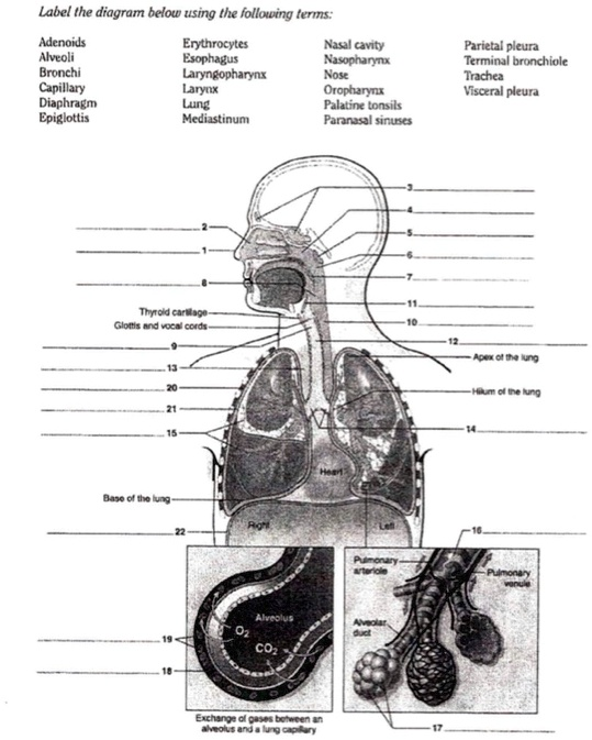
- Nose – external structure through which air enters
- Nasal cavity – air is filtered, warmed, and moistened
- Adenoids – lymphatic tissue in the upper nasopharynx
- Nasopharynx – upper portion of the pharynx
- Palatine tonsil – lymphoid tissue in the oropharynx
- Oropharynx – middle section of the pharynx
- Laryngopharynx – lower pharyngeal section before the esophagus and larynx
- Epiglottis – flap that prevents food from entering the airway
- Glottis – opening between the vocal cords in the larynx
- Larynx – voice box containing vocal cords
- Trachea – windpipe that leads to the bronchi
- Thread-like cartilage – refers to tracheal cartilage rings
- Bronchi – major air passages that branch from the trachea
- Terminal bronchiole – smallest airway before alveolar ducts
- Hilar pleura – pleura at the lung hilum
- Parietal pleura – outer membrane lining the thoracic cavity
- Lung – primary respiratory organ
- Mediastinum – central thoracic compartment between the lungs
- Alveoli – tiny sacs for gas exchange
- Capillary – blood vessels surrounding alveoli
- Erythrocytes – red blood cells
- Diaphragm – muscle enabling breathing
- Esophagus – muscular tube behind the trachea
- Adductor muscles – not part of respiratory anatomy; often relate to limbs
- Reticular layer – part of the dermis
- Dura mater – tough outer brain covering
- Meninges – collective brain coverings
- Tactile corpuscle – sensory receptor in skin
- Duct – a general term for channels transporting fluids
Explanation:
This diagram offers a detailed look at the respiratory system with both anatomical and microscopic structures. The upper airways begin with the nose and nasal cavity, which humidify and filter inhaled air. Lymphatic structures like adenoids and palatine tonsils aid in immune defense. Air passes through the pharynx (divided into nasopharynx, oropharynx, and laryngopharynx), then to the larynx, protected during swallowing by the epiglottis.
From there, it travels down the trachea, supported by thread-like cartilage, then enters the bronchi and progressively narrower branches like terminal bronchioles. Air reaches the alveoli, which are surrounded by a dense network of capillaries. Erythrocytes in these capillaries pick up oxygen and release carbon dioxide. The lungs are encased by the parietal and hilar pleura, within the thoracic cavity, separated by the mediastinum. The diaphragm contracts to create negative pressure that draws air in.
Though not part of the respiratory system, terms like reticular layer, duct, meninges, dura mater, and tactile corpuscle relate to skin and neural anatomy and are occasionally included in broader anatomical illustrations. This comprehensive labeling bridges macroscopic and microscopic views of human anatomy.