Drag and Drop 1 Articular cartilage 2 Proximal epiphysis 3 Diaphysis 4 Spongy bone 5 Distal epiphysis 6 Periosteum 7 Endosteum 8 Medullary cavity 9 Red bone marrow 10 Artery 11 Compact bone tissue 12 Yellow bone marrow
The Correct Answer and Explanation is:
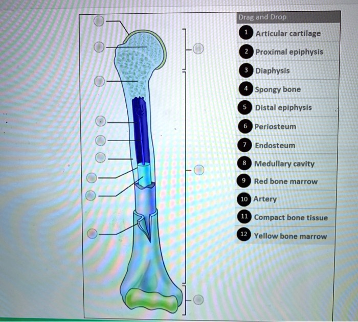
Here is the correct labeling for the parts of the long bone:
Labels on the Left Side (from top to bottom):
- Top label: 1. Articular cartilage
- Second label: 4. Spongy bone
- Third label: 6. Periosteum
- Fourth label: 8. Medullary cavity
- Fifth label: 7. Endosteum
- Sixth label: 10. Artery
- Seventh label: 12. Yellow bone marrow
- Eighth label: 11. Compact bone tissue
Labels on the Right Side (from top to bottom):
- Top label: 2. Proximal epiphysis
- Middle label: 3. Diaphysis
- Bottom label: 5. Distal epiphysis
Explanation of Long Bone Anatomy
This diagram illustrates the key structures of a typical long bone, which functions in support, movement, and blood cell production. The bone is divided into three main regions. The ends of the bone are the epiphyses. The proximal epiphysis (2) is the end closest to the body’s trunk, while the distal epiphysis (5) is the end farther away. The joint surfaces of the epiphyses are covered with articular cartilage (1), a smooth, resilient tissue that reduces friction and absorbs shock during movement. Internally, the epiphyses are filled with spongy bone (4), a porous, lightweight tissue whose spaces contain red bone marrow, the site of blood cell formation.
The long, main shaft of the bone is called the diaphysis (3). It is constructed from a thick, dense outer layer of compact bone tissue (11), which provides the bone with its strength and rigidity to withstand mechanical stress. The diaphysis is covered by a tough, fibrous outer membrane called the periosteum (6). The periosteum is essential for bone growth, repair, and the attachment of muscles and ligaments.
At the center of the diaphysis is the medullary cavity (8), a hollow space. This cavity is lined by a thin membrane known as the endosteum (7), which contains bone-forming and bone-resorbing cells. In adults, the medullary cavity is filled with yellow bone marrow (12), which is primarily composed of adipose tissue for energy storage. A nutrient artery (10) passes through the compact bone and runs within the medullary cavity to supply blood and nutrients to the bone tissue and marrow. Together, these components create a structure that is both strong and relatively lightweight.thumb_upthumb_down