Correctly label the anatomical features of the nose. Dense connective tissue Septal nasal cartilage Major alar cartilages Nasal bone Minor alar cartilages Lateral cartilage
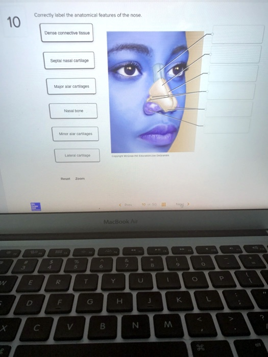
The Correct Answer and Explanation is:
Here is the correct labeling for the anatomical features of the external nose based on standard anatomical diagrams:
- Nasal bone – located at the superior part of the nose, forming the bridge
- Lateral cartilage – positioned just inferior and lateral to the nasal bone
- Septal nasal cartilage – forms the central wall dividing the nostrils
- Major alar cartilages – make up the rounded shape of the nostrils, forming most of the tip of the nose
- Minor alar cartilages – small cartilaginous structures that support the lateral portions of the nostrils
- Dense connective tissue – found at the apex of the nose, particularly around the nostrils where cartilage is absent
The external nose is a structural composite designed to balance rigidity with flexibility, allowing for effective airflow, olfaction, and facial expression. The nasal bones are paired structures that form the upper bridge of the nose, providing a rigid foundation. Inferior to these bones are the lateral cartilages, which extend the contour of the nasal sidewalls and support the midsection of the nose.
At the midline lies the septal nasal cartilage, a flat cartilaginous plate that divides the nasal cavity into right and left halves. This septum also anchors adjacent cartilages and maintains structural integrity.
The nostrils are shaped and supported by the major alar cartilages, which form a curved structure creating the tip and medial walls of the nares. Additional support comes from minor alar cartilages, located laterally and often varying in number. These help maintain the patency and shape of the nostril openings.
At the most inferior and anterior part of the nose, dense connective tissue replaces cartilage where structural rigidity is less necessary. This tissue allows for flexibility while still offering mechanical strength to resist deformation.
Understanding this anatomy is essential for assessing trauma, planning surgeries like rhinoplasty, and diagnosing congenital or acquired nasal deformities.