Activity 3: Dissecting the Mammalian Eye 1. Identify the labeled structures in the accompanying photographs of a dissected cow eye: a. b. c. d. e. Activity 4: Performing Visual Tests
The Correct Answer and Explanation is:
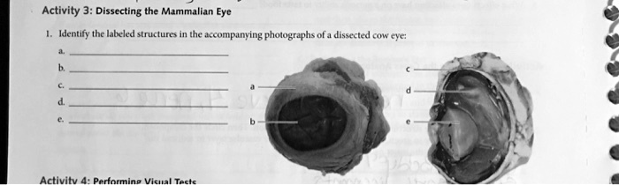
Based on the provided images of a dissected cow eye, here are the identified structures:
a. Iris
b. Pupil
c. Retina
d. Tapetum lucidum
e. Lens
Explanation
The photographs show two different views of a dissected cow eye to reveal its key anatomical features. The image on the left provides an anterior view, looking into the front of the eye, while the image on the right displays a sagittal cross section, showing the internal layers and structures.
The structure labeled a is the iris. The iris is the pigmented, muscular diaphragm that surrounds the pupil. Its function is to control the size of the pupil, thereby regulating the amount of light that enters the eye and reaches the retina.
The opening in the center of the iris is the pupil, labeled as b. Light passes through the transparent cornea and then through the pupil to get to the lens. In cows and many other grazing animals, the pupil is not round like in humans but is horizontally oval, which provides a wider panoramic field of view.
In the cross section view, the structure labeled c is the retina. This is the innermost, light sensitive layer of tissue at the back of the eye. The retina contains photoreceptor cells, known as rods and cones, which detect light and convert it into neural signals. These signals are then sent to the brain through the optic nerve for visual processing.
Label d points to the tapetum lucidum. This is a reflective, often iridescent layer located just behind the retina in the choroid layer. It is a feature common in nocturnal or crepuscular animals like cows but is not present in human eyes. The tapetum lucidum reflects light back through the retina, giving the photoreceptors a second opportunity to capture photons. This significantly enhances night vision and is responsible for the “eyeshine” effect when light is shone into the animal’s eyes in the dark.
Finally, the structure labeled e is the lens. The lens is a transparent, biconvex structure located directly behind the iris and pupil. Its primary function is to focus light onto the retina, creating a clear, sharp image. The lens can change its curvature to adjust focus for objects at different distances. After dissection and preservation, the lens often becomes hard and opaque.