Art-labeling Activity: The Structure of a Lymph Node Part A Rest Hope
The Correct Answer and Explanation is:
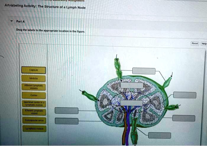
ere is the correctly labeled image of the structure of a lymph node.
Explanation of the Lymph Node Structure
A lymph node is a crucial secondary lymphoid organ that functions as a sophisticated filter for lymph, trapping pathogens and initiating adaptive immune responses. Its structure is highly organized to facilitate this role.
The entire node is enclosed by a dense fibrous Capsule, which also extends inward as trabeculae, dividing the node into compartments. Lymph fluid enters the node through several Afferent lymphatic vessels located on its convex surface. From there, the lymph flows into the Subcapsular sinus, a space directly beneath the capsule.
The lymph then percolates through the node’s outer region, the Cortex. The cortex is populated by B lymphocytes organized into distinct spherical structures called Lymphatic nodules or follicles. When these nodules are actively responding to an antigen, they develop a lighter-staining central region known as a Germinal center in a lymphatic nodule. This area is a site of intense B cell proliferation and differentiation into antibody producing plasma cells.
After filtering through the cortex, lymph moves into the central Medulla. The medulla contains cords of lymphatic tissue with T cells, B cells, and plasma cells. The lymph slowly flows through medullary sinuses, allowing for further interaction with immune cells.
Finally, the filtered lymph is collected and exits the lymph node at its indented region, the hilum, through one or two Efferent lymphatic vessels. This one way flow of lymph through the node ensures maximum exposure to lymphocytes and macrophages, making the lymph node an effective site for immune surveillance and the activation of the body’s defenses against infection.