Identify the parts of an osteon in the photomicrograph b d. a. b. c. d.
The Correct Answer and Explanation is:
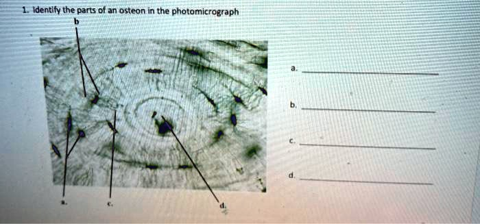
Based on the photomicrograph of the osteon, here are the identified parts:
a. Lacuna (plural: lacunae)
b. Canaliculus (plural: canaliculi)
c. Lamella (plural: lamellae)
d. Central canal (Haversian canal)
Explanation
The photomicrograph displays a cross section of compact bone, showing its fundamental structural unit, the osteon or Haversian system. Each component labeled is essential for the function and integrity of bone tissue.
The structure labeled d is the central canal, also known as the Haversian canal. This channel runs longitudinally through the center of the osteon. It is a critical passageway that houses blood vessels, nerves, and lymphatic vessels. This vascular network is responsible for supplying the bone cells with oxygen and nutrients while also removing metabolic waste products, ensuring the tissue remains alive and healthy.
Surrounding the central canal are the concentric rings of calcified bone matrix, identified as c, the lamellae. These layers give compact bone its immense strength. The lamellae are composed of collagen fibers and inorganic mineral salts, primarily calcium phosphate. The collagen fibers within each lamella are arranged in a parallel fashion, but the orientation of fibers in adjacent lamellae alternates. This alternating pattern allows the osteon to effectively resist twisting forces.
Embedded within the lamellae are small cavities called lacunae, labeled as a. Each lacuna is the space that houses a mature bone cell called an osteocyte. These osteocytes are responsible for maintaining the surrounding bone matrix and sensing mechanical stress.
Radiating from each lacuna are tiny, hair like channels known as canaliculi, labeled as b. These microscopic canals form an intricate network connecting the lacunae to one another and to the central canal. The cytoplasmic processes of the osteocytes extend through these canaliculi, forming gap junctions with neighboring cells. This network is vital for communication and for the transport of nutrients and wastes between the blood vessels in the central canal and the osteocytes trapped within the hard matrix, thus keeping the bone cells viable.